Sperm smear histology


To examine sperm morphology, prepare at least two smears after the semen has liquified (>30 minutes). The specimen should be thoroughly mixed before.


Sperm Smear Histology - an album on Flickr
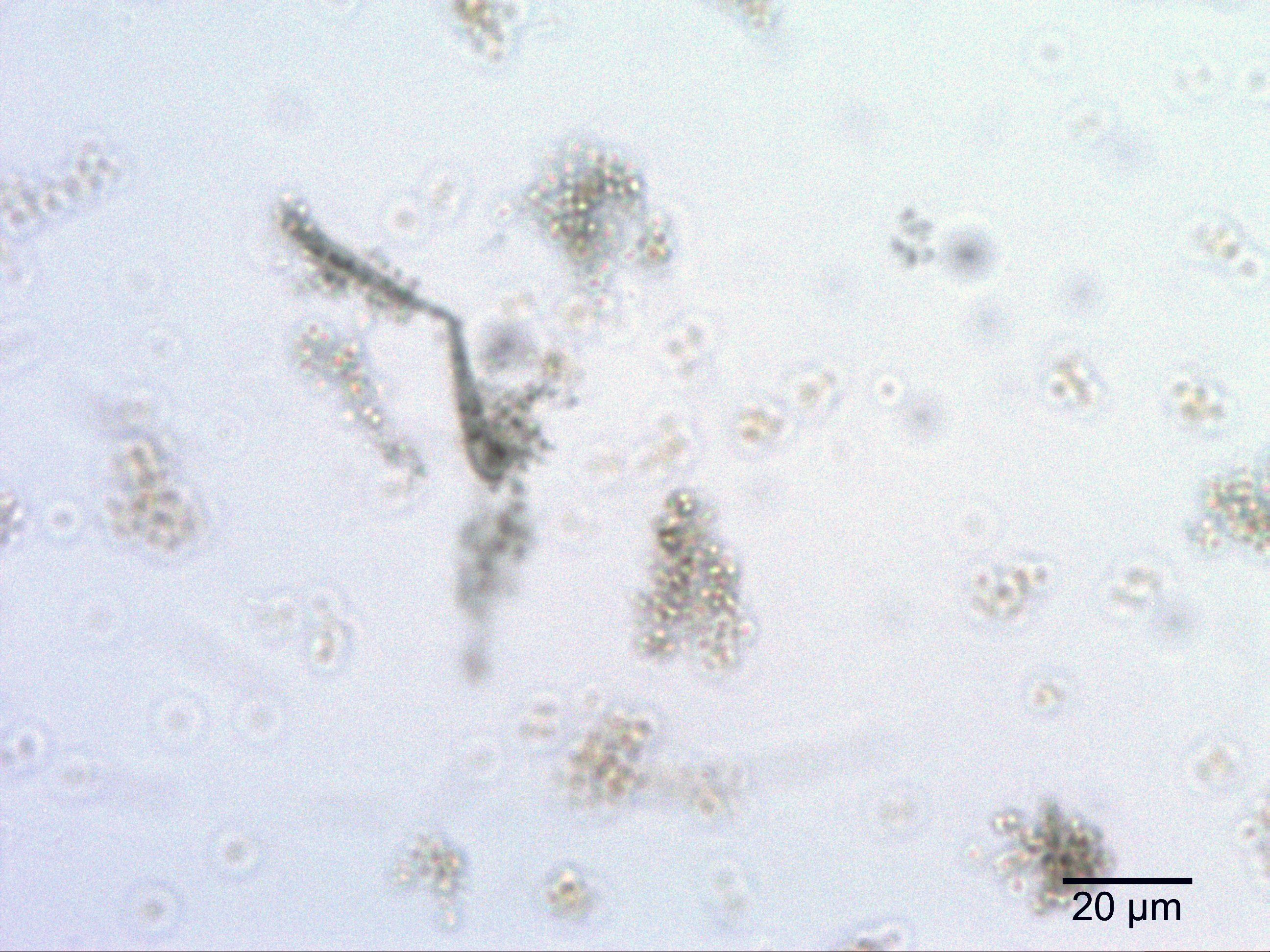
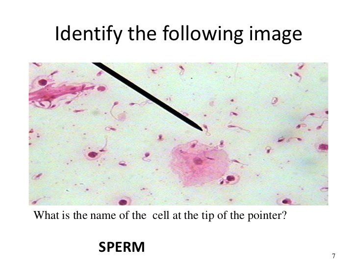
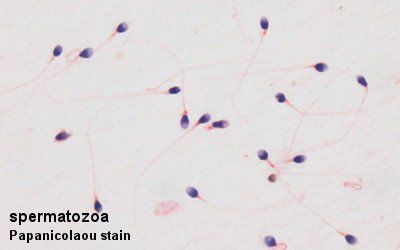
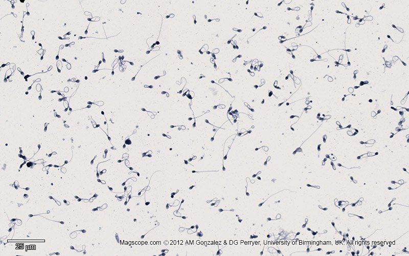
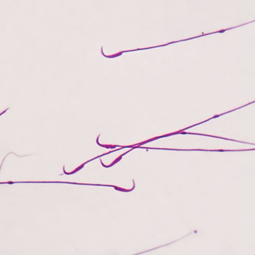
Mammal Bull Sperm, smear Microscope Slide
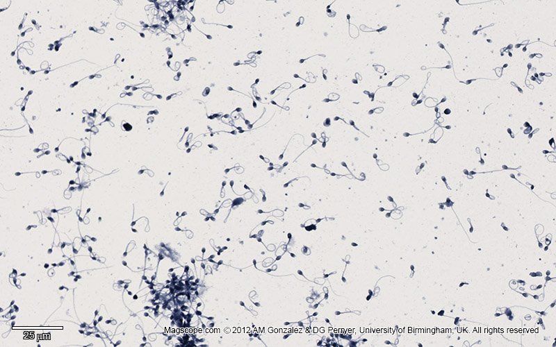
In the first part of the study, smears were stained with Hematoxylin Eosin (HE), Toluidin Blue KEY WORDS: Spermatozoa; Morphology; Histological stains.

I Kimberley Age: 34. Bises,sex, normal sex in any position you want erotic massage.Je propose une rencontre de qualitй adaptйe a des gentlemen exigeants et sйlectifs.-Leidenschaftliche Zweisamkeit
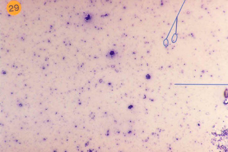
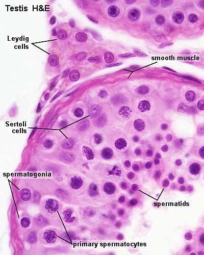
Sperm smear, human
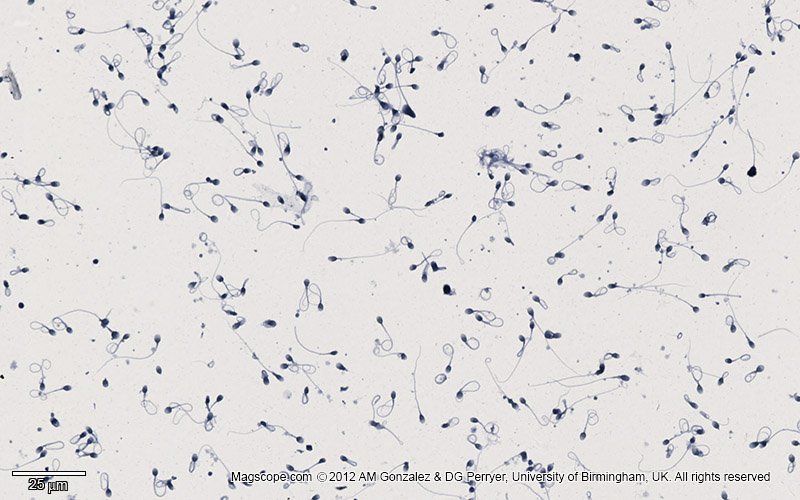


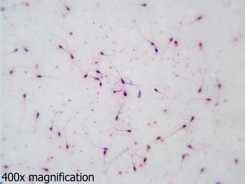
Histology Slides for Urinary System Sperm Smear/human Teaching Supplies: Biology Classroom:Biology Classroom Microscope Slides.

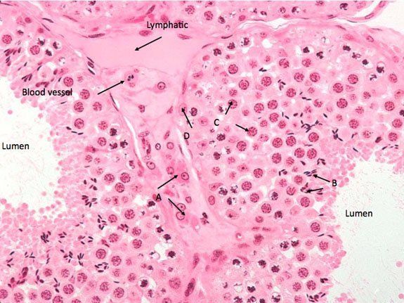
Description:Tight junctions may temporarily open to permit the passage of spermatogenic cells from the basal into the adluminal compartment. A fold in the nuclear membrane is characteristic for Sertoli cells but not always visible in the LM well Sertoli cells are far less numerous than the spermatogenic cells and are evenly distributed between them. They appear larger than spermatogonia. An nigrosin-eosin stain is commonly used because it is effective, simple and, in addition to allowing sperm to be readily visualized, it is a so-called "live-dead" stain, allowing one to assess membrane integrity at the same time as morphology. Examine using a bright field microscope typically using a X objective lens. The tunica albuginea is covered externally by a serosa.


User Comments 4
Post a comment
Comment: